Let’s look at what happens when we use a 180 pulse instead of a 90. Now, the spin is pointed straight down instead of in the MXY plane.
Here is the final move, and it’s a favorite: put a 180° inversion pulse in front of everything. Instead of knocking the longitudinal magnetization down to zero, it flips it all the way to −M0. From there it recovers up through zero and back toward +M0, following almost the same curve as before but starting from the bottom:
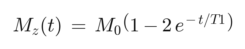
Because it starts negative and climbs, every tissue’s curve must pass through zero on the way up. Set the exponential equal to the crossing point and the “null time” falls right out:

`TI` (inversion time) is how long we wait after the inversion pulse before we excite and read out. If we set TI to a tissue’s null point, that tissue has zero longitudinal signal at that instant and contributes nothing to the image — effectively “disappearing” since we suppressed. And since the null time depends on T1, different tissues null at different TIs:
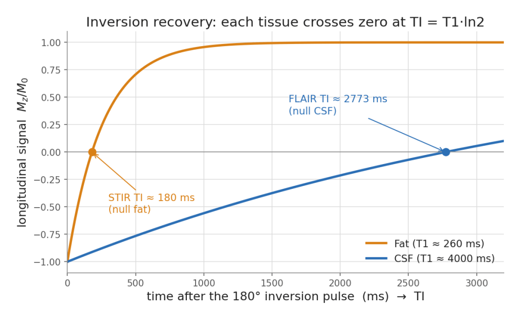
– STIR — pick the short TI that nulls **fat**. Because it nulls by T1 rather than by fat’s precession frequency, STIR suppresses fat robustly even where the field is uneven — handy off-isocenter and in the extremities.
– FLAIR — pick the long TI that nulls **CSF**, so bright fluid stops drowning out periventricular disease. A FLAIR makes an multiple sclerosis (MS0 plaque or a rim of edema pop next to a now-dark ventricle.
Be careful here – STIR and FLAIR imply different TIs – but are essentially the same sequence.
At the PACS station, that is the entire logic: which tissue did we choose to send through zero at readout?
And that closes the loop. With one signal equation, two knobs (TR and TE), the two relaxation times behind them, the echo that measures them, the flip angle that tunes them, and two ways to add contrast (gadolinium and inversion), you can reason your way through almost any sequence card on the scanner.
 Carolina Laboratory for Advanced Radiological Imaging and Technology