TR controls the recovery of the longitudinal signal. TE controls the opposite process happening to the signal we actually measure — the transverse part. We also adjust TE to control contrast.
The instant after excitation, the transverse magnetization `Mxy` (the part we can detect) is at its maximum, and from there it only shrinks. It decays exponentially with a time constant called T2:
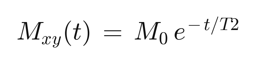
There is our `e^(−TE/T2)` term again. **TE is how long we wait after excitation before we read the signal out** — how far down this decay curve each tissue has slid when we measure it.
Plot the decay for a long-T2 tissue (fluid) and a short-T2 tissue (most soft tissue), and drop a vertical line at a long TE:
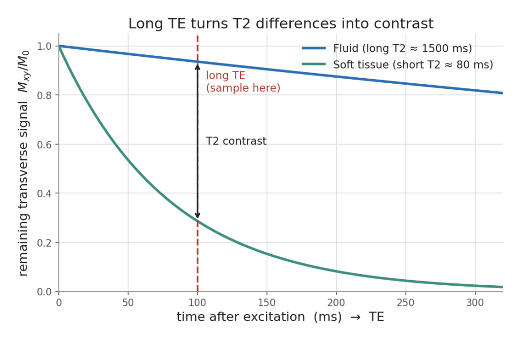
At a short TE (far left) both tissues still have most of their signal and look similar — that is why the T1 and PD images used a short TE to hide T2 differences.
Wait until a long TE and the short-T2 tissue has faded to almost nothing while the fluid is still bright. That gap is your T2 contrast, and it is why fluid lights up on T2.
Two knobs, two exponential curves: TR walks each tissue up its recovery curve, TE walks it down its decay curve, and where you choose to sample decides the picture.
We have leaned hard on the idea that fat has a short T1 and water a long T1 and T2.
Why should that be?
 Carolina Laboratory for Advanced Radiological Imaging and Technology